Ola Abdalsalam has developed a novel method to enhance the accuracy of brain activity measurements in infants. This new approach, called structured interrogation (SI), uses advanced light-based technology to peer deeper into tissues, offering improvements over traditional techniques.
Key Findings:
-
Structured Interrogation (SI) Technique:
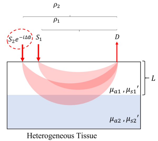
- SI utilizes light waves that interfere with each other by adjusting the phase of their modulation. This allows for a more precise examination of tissues, enhancing the clarity and detail of the images produced.
- Unlike traditional methods, SI can dynamically adjust without the need for extra light sources or photodetectors, making it more efficient and versatile..
-
Improved Sensitivity:
- The phase-only component of SI FD-NIRS (Frequency-Domain Near-Infrared Spectroscopy) can accurately measure both absorption and scattering of light in tissues.
- SI shows a 20% improvement in detecting changes in deeper tissues compared to conventional methods.
-
Application to Functional Brain Monitoring:
- This technique is particularly promising for monitoring brain activity in infants. It reduces interference from superficial layers like the scalp and skull, leading to clearer and more accurate readings of brain activity.
- Enhanced sensitivity provided by SI could improve how we understand and noninvasively monitor brain function in early development stages.
-
Validation and Performance:
- Using tissue-like models, SI demonstrated less than 5% error in estimating tissue properties, even in the presence of noise.
- The method was rigorously tested on both single-layer and multi-layer tissue models, proving its robustness and effectiveness across different scenarios.
This innovative technique offers a step forward in noninvasive medical imaging, with potential applications extending beyond brain monitoring to other areas requiring detailed tissue analysis such as breast cancer detection and monitoring.
For further details on the study, please refer to the full publication in the Journal of the Optical Society of America A, August 2024 issue.